The ability to locate and visualize proteins and macromolecular complexes in cells and tissues in 3D high resolution continues to be a challenge in biomedical studies. Various techniques and tools are key to this work. For example, light microscopy uses fluorescent labels to track elements of interest, but it provides an incomplete view, including only the distribution of the labeled elements. Researchers who want to collect more comprehensive 3D datasets at higher-resolution use electron microscopic tomography (EMT) to visualize all structures in the cellular or subcellular domain under investigation. However, EMT remains a relatively complicated method. To obtain 3D reconstructions of biological samples (tissue or cell monolayer), which are typically embedded in plastic, semi-thin sections must be sliced by ultramicrotomy, then imaged at different sample orientations, yielding a comprehensive set of 2D digital electron micrographs. These micrographs are then processed through a workflow to create the final 3D volume for analysis.
The National Center for Microscopy and Imaging Research (NCMIR) has long been known for its state-of- the-art work in EMT, in particular developing a framework designed to correct for imperfections due to electron microscope optics and distortions arising from the sample warping. All this enables fine 3D reconstructions of biological specimens.
In collaboration with the National Biomedical Computation Resoruce (NIGMS P41), the NCMIR team has developed a novel automated method for multi-tilt data acquisition allowing for a greater number of views of a sample of interest at many different orientations than has been possible in the past. This is achievable because acquiring a large amount of data for plastic-embedded samples, does not affect the sample integrity as long as the electron beam dose-rate is maintained low. The reconstruction space can be sampled in a more refined way with respect to traditional methods, leading to improved quality in the 3D representation. This methodology makes it easier to visualize and analyze the details of biological structures around the nanometer scale. In fact, by accumulating large numbers of lower-dose electron micrographs, the contrast in the final reconstruction is greatly enhanced such that mild staining protocols are sufficient, and allows distinguishing more molecular details.
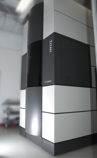
In contrast to the traditional single- and double-tilt- series acquisition protocol, this approach requires orienting the sample within the electron beam with a more complex scheme involving wider range of specimen orientations. In the work described here the team used a data-acquisition scheme of 16 tilts. Up to 2,000 micrographs were obtained using an FEI Titan 300-kV microscope outfitted with a 4k x 4k Gatan charge-coupled device camera. By averaging a greater number of tilt series together, artifacts that would normally hamper the reconstruction are reduced as the rays crossing the sample at any point are more evenly distributed from an angular perspective. To reduce the additional complexity of this strategy, the team developed automated procedures for alignment and volume-generation processes, reducing (and even eliminating) the requirement for user input.
In their work the NCMIR/NBCR team applied this method to reconstructions of two biological structures in situ in their normal intracellular contexts: the double-stranded giant DNA Mimivirus and clathrin-coated vesicles, which transport cargo such as proteins between organelles in eukaryotic cells. Both have been studied extensively by other methods, providing useful reference data on the details of their structures with which to compare the results obtained using this multi-tilt EMT approach.
Sections from both sample types were cut using a diamond knife at a thickness of 200- 300nm. Gold particles (5 and 10nm in diameter) were deposited on each side of the sections to serve as fiducial markers. These markers provide a way to track areas of interest across the sections and align the 2D digital electron micrographs precisely into a 3D volume
The images were aligned using TxBR, a software package developed at NCMIR that optimizes micrograph registration to the final volume using the gold fiducial markers. TxBR accounts for the curvilinear nature of the electron trajectories in the microscope, making it well suited to processing wide-field electron microscope data, and enables 3D reconstruction from various data-acquisition schemes, including any number of tilt series as shown in this work. TxBR is able to correct for considerable sample warping or deformation, optical distortion, and non-linear trajectories of the electron beam. In their work, the team used and compared two methods to reconstruct the 3D volume: a simple filtered back projection scheme, and a more elaborated iterative procedure. High-quality reconstructions were obtained with both procedures with slight improvements for the latter. However, in a high-throughput EMT production environment, implementing the back projection scheme might be advantageous because it is less computer intensive. This leads to a practical tradeoff: Should one acquire a smaller number of tilt series (2-4) with the iterative approach, or a larger number of tilt series and restrict the reconstruction process to filtered backprojections. Additional investigations are currently being performed.
Funding Source:
This work was conducted at the National Center for Microscopy and Imaging Research in collaboration with the National Biomedical Computation Resource. NCMIR and NBCR are NIGMS P41 Research Resources supported by NIH Grants GM103412 and GM103426 awarded to Dr. Mark Ellisman and Dr. Rommie Amaro, respectively.
Relevant Publication:
Phan, S., Boassa, D., Nguyen, P., Wan, X., Lanman, J., Lawrence, A., Ellisman, M. H., (2017), “3D reconstruction of biological structures: automated procedures for alignment and reconstruction of multiple tilt series in electron tomography”, Adv Struct Chem Imaging, 2016/08/23, 2: pg: 8, 2198-0926, (DOI: 10.1186/s40679-016- 0021-2).